
11 Jan Diagnosing & Treating Melanoma: A Quinn Dermatology Case Study
Melanoma Diagnosis
A white man in his late 60’s came in for a skin evaluation over the summer of 2023. After being referred by his primary care physician, he presented with a suspicious lesion on his face that had been growing slowly for about three months. Upon a full skin exam, an incidental finding of a lesion on his back was discovered, which was a growth that the patient had not been aware of.
While melanoma often appears initially like a harmless mole, recent studies show that most melanomas (an estimated 70 percent) start as a brand-new lesion, not an existing mole (often referred to as “de novo” lesions).
The fact that this patient presented for a benign growth on his face that turned out to be seborrheic keratosis, highlights the importance of routine skin exams as the cancer would not have been found if he had not presented for this unrelated lesion.
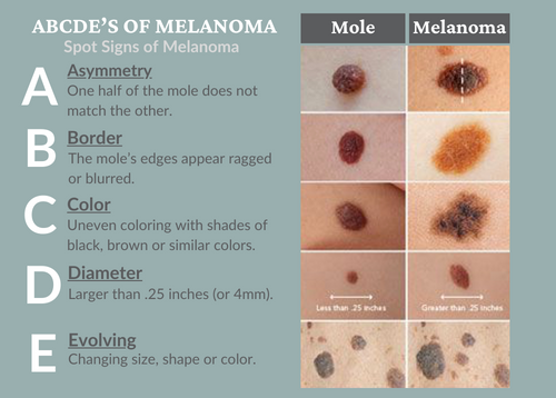
When diagnosing melanoma, Dr. Quinn assesses his patient’s entire body for suspicious lesions, using the ABCDE evaluation method: A for asymmetry, B for border, C for color (variation), D for diameter (greater than 5 mm), and E for evolving/erythema.
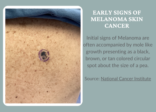
When evaluating this patient, Dr. Quinn found that the growth on the back presented as a singular, 1.6cm black and brown plaque with ill-defined borders, which was immediately biopsied and later found to be an invasive melanoma, an aggressive and at times life threatening skin cancer.
Before and after confirming the diagnosis with the biopsy, the patient was brought back for a wide local excision that removed the scar where the tumor was found along with 1cm of surrounding normal skin down to the muscle.
Melanoma Treatment
The stage of the cancer determines what treatment options to consider. In this case, the biopsy results showed stage IA, which generally only requires surgery at the site the tumor is found using a scalpel to cut away the entire mole and some of the healthy tissue around it.
Melanoma stages use the numbers 0 through 4. At stage 0 and stage 1, a melanoma is thin and small. Treatment is likely to be successful. As the melanoma grows deeper into the skin, the stages get higher. Treatment becomes more challenging. By stage 4, the cancer has spread beyond the skin to other organs, such as the lungs or liver.
Immediate post treatment included a three-month post-surgery evaluation with visual and tactile assessments and an interview questioning the patient about possible symptoms. The treated area was examined visually with the naked eye and with a dermascope. Additionally, lymph node basins were palpated to screen for lymphadenopathy (enlargement of lymph nodes), to screen for metastasis. In this case, the patient checked out fine and did not require any further immediate assessments.
Final Case Outcome
The patient experienced no complications after surgical excision. He is expected to make a full recovery with a 7cm linear scar. The patient was educated to conduct skin self-examinations, to use sunscreen when outdoors, and to return every 3 months for monitoring and total body skin examinations.